Prostate MRI vs Biopsy: How advanced imaging is reducing unnecessary tests
By Dr Marcus Stephan • Published: 01 Sep 2026 • 9:00 • 6 minutes read
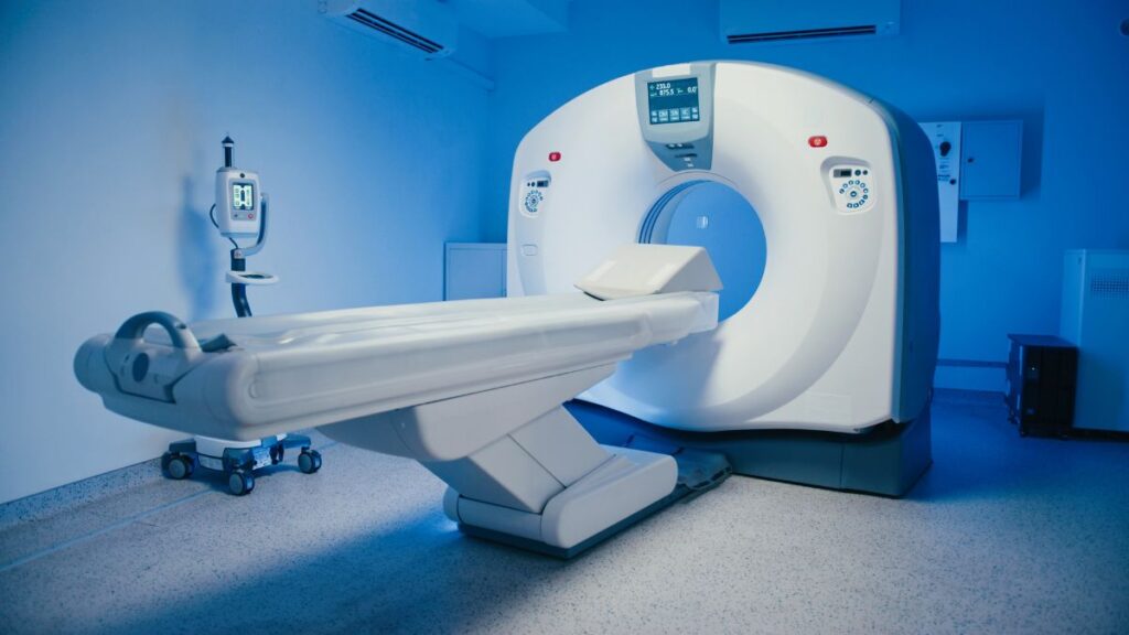
Advanced multiparametric MRI scans help doctors determine whether an invasive prostate biopsy is necessary. Credit: VesnaArt/Shutterstock
Medical science has reached a major turning point, particularly in how we approach early cancer detection. For decades, diagnosing prostate cancer followed a single, invasive principle: if cancer was suspected, a needle biopsy was performed to examine tissue under a microscope. Today, multiparametric magnetic resonance imaging (MRI) is reshaping that traditional pathway. Under updated 2026 European Association of Urology (EAU) guidelines, a reassuring MRI scan can help thousands of men safely avoid an unnecessary biopsy altogether.
This year, medical journals are full of some really fascinating breakthroughs – it really feels like medical science it turning a major corner; from new ‘personal care’ where drugs are designed for you, to some really exciting advances in the way we treat cancers.
I have written about Prostate Cancer before and make no apologies for returning to the subject. This cancer is very treatable if caught early, however for many men the prospect of a biopsy raises real, even if fairly remote, chances of risk. For decades, the diagnosis of prostate cancer has followed a fairly simple principle: if cancer is suspected, take a piece of the prostate and examine it under a microscope. The biopsy has traditionally been regarded as the definitive test.
That position is now changing.
Magnetic resonance imaging (MRI), particularly the sophisticated form known as multiparametric MRI, has become an increasingly important part of prostate cancer diagnosis. The latest 2026 European guidelines go further than before in recognising that, for carefully selected men, a reassuring MRI can mean that a biopsy may safely be avoided completely.
But there is an important distinction. MRI has not replaced biopsy as the definitive means of proving that prostate cancer is present. Instead, MRI has become an extremely powerful tool for deciding whether a biopsy is even necessary.
Why conventional prostate biopsies carry hidden risks
A conventional prostate biopsy involves inserting a needle into the prostate and removing a number of tiny cores of tissue. A pathologist then examines those cores under a microscope, looking for malignant cells and determining how aggressive the cancer appears to be.
This microscopic examination provides information that an MRI cannot currently provide with the same certainty. The pathologist can determine the tumour’s grade, including the internationally recognised Gleason and ISUP Grade Group systems. That information is crucial when deciding whether a cancer requires treatment or can safely be monitored.
Biopsy, however, is far from perfect.
It is invasive, can cause bleeding and infection and, particularly with older transrectal techniques, carries a small but potentially serious risk of sepsis. It can also find cancers that are technically present but so slow-growing that they would never have caused any problems.
Multiparametric MRI and the PI-RADS Scoring System
Multiparametric MRI provides remarkably detailed images of the prostate. It combines several different types of MRI imaging to identify areas that look suspicious for clinically significant cancer.
Radiologists generally report these findings using the PI-RADS system, which grades abnormalities from 1 to 5. A PI-RADS 1 or 2 scan is considered unlikely to contain clinically significant cancer. PI-RADS 3 is indeterminate, while PI-RADS 4 or 5 indicates an increasing likelihood of significant cancer.
The crucial development is that doctors no longer have to regard the MRI simply as a way of finding a target for the biopsy.
They can use it as part of the decision about whether the biopsy should happen at all.
What the 2026 EAU Guidelines say about pre-biopsy scans
The 2026 EAU guidelines recommend performing MRI before biopsy in men with suspected cancer confined to the prostate. They also specifically recommend using multiparametric MRI to avoid unnecessary biopsies. For a man with a negative MRI and a low overall risk of significant cancer, the guidelines say that biopsy can be omitted and PSA monitoring offered instead.
That is a significant change in philosophy.
How reliable is a negative MRI scan result?
This is where the story becomes particularly interesting.
A large body of evidence now shows that a good-quality MRI has a high negative predictive value for clinically significant prostate cancer, particularly when combined with a low PSA density.
In plain English, a reassuring MRI combined with reassuring clinical information makes the likelihood of an important cancer considerably smaller.
But “smaller” does not mean “zero”.
Some significant cancers are invisible or poorly visible on MRI. A negative MRI can therefore never be interpreted as an absolute guarantee that cancer is absent.
This is why the latest guidance still stresses clinical judgement and follow-up.
Precision diagnosis: The benefits of MRI-targeted biopsies
This is where MRI has perhaps made the biggest difference.
If the MRI shows a suspicious lesion, the radiologist can identify its precise location. The biopsy needle can then be directed specifically towards that area.
This is known as an MRI-targeted biopsy.
Rather than taking numerous samples more or less blindly from different parts of the prostate, the clinician can concentrate on the suspicious area, often combining targeted samples with a smaller number of regional or systematic samples. The 2026 EAU guidelines now recommend targeted and regional sampling when a biopsy is undertaken.
This approach is considerably more sophisticated than the traditional “needle sampling” method.
Can an MRI imaging scan confirm cancer without a biopsy?
Not quite — and this is the point that is sometimes lost in some media reports.
The 2026 EAU guidelines state that definitive diagnosis normally depends on histopathological verification — in other words, examination of prostate tissue obtained by biopsy. MRI can show that an area looks extremely suspicious for cancer. It can even provide a very strong indication that cancer is present.
But an MRI image cannot, by itself, demonstrate malignant cells under a microscope.
There are exceptions. In a man with overwhelming evidence of advanced prostate cancer — for example, a very high PSA, a prostate that feels frankly malignant and evidence of metastatic disease — a biopsy may sometimes be unnecessary, particularly when the result would not alter treatment.
For the ordinary man being investigated for possible localised prostate cancer, however, biopsy remains the definitive confirmation.
Avoiding unnecessary biopsies: The real healthcare revolution
Perhaps the most important change is therefore not “MRI replaces biopsy”.
Rather MRI helps determine who actually needs a biopsy.
The EAU describes an MRI-based pathway in which men with a positive MRI undergo targeted biopsy while those with a negative MRI may avoid biopsy altogether. Depending on the criteria used, this approach could avoid biopsy in a substantial proportion of men, although a small number of clinically significant cancers will inevitably be missed.
The guidelines also report research in which a risk-based pathway allowed 19% of men to avoid biopsy while missing clinically significant cancer in approximately 1.2%.
That represents a very different approach from the traditional philosophy of “biopsy everyone who might have cancer”.
What the modern risk-based diagnosis pathway means for patients
The modern prostate cancer pathway is becoming a risk assessment rather than a simple sequence of tests.
A raised PSA does not automatically mean biopsy. The PSA result is considered alongside prostate size, PSA density, age, family history, examination findings and, increasingly, MRI.
A low-risk combination may justify monitoring rather than immediate biopsy. A suspicious MRI, on the other hand, can direct the biopsy precisely to the area that matters. And if the biopsy confirms cancer, the tissue provides information about the grade and biological characteristics of the tumour that MRI cannot currently replace.
So the biopsy is not disappearing.
It is becoming more selective.
That may ultimately be the greatest achievement of modern prostate MRI. Instead of using biopsy as the first major step towards a diagnosis, doctors can increasingly use MRI to decide who genuinely needs one.
For men with prostate cancer, that is an important distinction. It means fewer unnecessary invasive procedures, fewer diagnoses of harmless cancers and, when cancer really is present, a much better chance of targeting the biopsy at the disease that matters.
The future is therefore unlikely to be “MRI versus biopsy”. It is much more likely to be MRI first, biopsy when necessary.
And according to the latest European guidance, that future is already here.
The information provided in this column is for educational and informational purposes only, and does not constitute medical advice. It is not a substitute for a professional medical consultation, diagnosis, or treatment. Always seek the advice of your own physician or other qualified health provider with any questions you may have regarding a medical condition.
Dr Marcus Stephan
Follow Euro Weekly News on Google News
Get breaking news from Spain, travel updates, and expat stories directly on your Google News feed.
Follow on Google NewsSign up for personalised news
Subscribe to our Euro Weekly News alerts to get the latest stories into your inbox!
By signing up, you will create a Euro Weekly News account if you don't already have one. Review our Privacy Policy for more information about our privacy practices.





Dr Marcus Stephan
With a career spanning more than 35 years in both the UK and internationally, Dr Marcus' passion lies in empowering individuals through knowledge, enabling them to care for themselves and others. He simplifies the science behind medicine by removing unnecessary jargon, explains common health conditions, and shares updates on the latest advances and developments in medicine.
Comments